


Coronary angiography is performed to detect obstruction in the coronary arteries of the heart. During the procedure a catheter (thin flexible tube) is inserted into an artery in your arm or groin and then threaded carefully into the heart. The blood vessels of the heart are then studied by injection of contrast media through the catheter. A rapid succession of X-rays (fluoroscopy) is taken to view blood flow.

Coronary angioplasty is accomplished using a balloon-tipped catheter inserted through an artery in the groin or arm to dilate the coronary artery. Coronary artery disease occurs when cholesterol plaque builds up (atherosclerosis) in the walls of the arteries of the heart. Angioplasty is successful in opening coronary arteries in 90% of patients. 40% of patients with successful coronary angioplasty may develop recurrent narrowing at the site of balloon inflation.


Computed Tomography (CT or “CAT” Scan) scanning is a rapid, painless diagnostic examination that combines x-rays and computers. A CT scan allows the radiologist to see the location, nature, and extent of many different diseases or abnormalities inside your body. CT scanning can be used to obtain information about almost any body organ (such as the liver, pancreas, intestines, kidneys, adrenal glands, lungs, and heart), blood vessels, the abdominal cavity, bones, and the spinal cord. Our center has Spiral CT scanners equipped with new software techniques and workstations i.e. Advantage Workstation 4.2 which generate data and detail quickly and accurately.

Critical Care Unit (CCU) represents the apex of technical advancements in medicine. Such units integrate many specialties and diverse technologies, offering the possibility of survival to patients who would otherwise die. It is therefore obvious that the role of critical care services is vital and crucial to a tertiary care hospital, as the critical care unit contributes immensely to survival of acutely and critically ill patients


Digital (or computerized) imaging techniques came to x-ray in the 1980s when analog to digital (A/D) converters and computers were also adapted to conventional fluoroscopic image intensifier/TV systems. “Digital imaging” has lead to a similar improvement and renaissance in x-ray. Digital x-rays often look sharper and cleaner than the analog version. Many of the fluoroscopic (“fluoro” for short) x-ray procedures described herein have benefited greatly from the addition of digital technology. Further, angiographic procedures for looking at the blood vessels in the brain, kidneys, arms and legs, and the blood vessels of the heart all have benefited tremendously from the adaptation of digital technology. At SSMH has fully motorized Diagnostic X-Ray Table and X-Ray Machine is ideal for all routine and special examinations. FUJI Computer aided digital X-Ray with laser Camera for post processing and reduction/ magnificent software for high quality world class imaging.

-Facility-1657894724.jpg)


To give a new lease of life to the critically ill, cardiac patients – SSMH houses an Intensive Coronary Care Unit (ICCU) with 12 beds, providing advanced monitoring and life support systems under supervision of capable cardiologist & attending resident doctors. We also house a 10 bedded High Dependency Unit (HDU) for seriously ill patients, who need specialist nursing care and monitoring.

-1657796163.jpg)
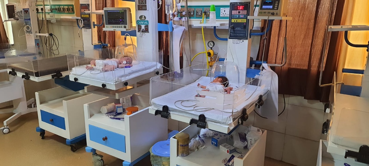
The Paediatric care in SSMH extends from birth to adolescence. There is a 10 bedded well spaced aseptic nursery for low risk babies, with facilities of Phototherapy, radiant warmers, infusion pumps, central oxygen and suction etc. For high risk and sick babies, there is a full fledged Neonatal Intensive Care Unit (NICU) with CPAP, ventilators and proper nursing care.


Ultrasound is a simple, safe, painless diagnostic procedure that bounces high-frequency sound waves off parts of the body and captures the returning “echoes” as images. There is no injection or radiation exposure associated with ultrasound. SSMH offers many different types of ultrasound exams. Ultrasound is able to capture moving images of pelvic and abdominal function (including gallstones), breast abnormalities, the male reproductive system, the kidney and thyroid systems, as well as the developing fetus, among other applications. When enhanced with a special Doppler technique, ultrasound can also capture moving blood images of large blood vessels and moving images of the heart using echocardiography.
